Dynamics of Complex Biological Systems

At the convergence of physics with biology, our group is motivated both by the desire to gain fundamental insights into the behavior of living systems and to contribute to the pressing challenges associated with the explosion of quantitative information in medical research.

Our analysis of shape dynamics of migrating cells has led us to discover mechanical waves as a ubiquitous underlying motors in many fast-migrating cells. Another project in the Biodynamics lab is to elucidate how surface chemistry and topography affects migratory machinery, and how internal waves may be harnessed to control cell behavior. We also developed new tools to control the arrangement and dynamics of cell groups via holographic laser tweezers and to investigate the mechanical properties of models of circulating tumor cells. Other projects apply Complex Systems approaches to investigate cancer related biological processes as part of a Cancer Technology interaction between the University of Maryland and the National Cancer Institute. Examples of the many ways studying complex systems provides insight into biological systems are available on this page.

Collective Migration during Cancer Progression

Rachel Lee, Postdoctoral Research Associate
with Dr. Stuart Martin (University of Maryland School of Medicine)

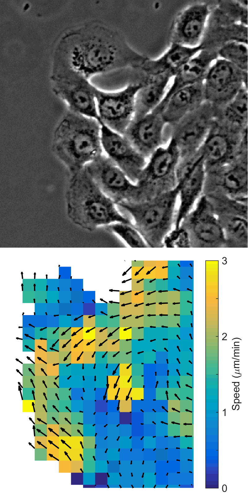
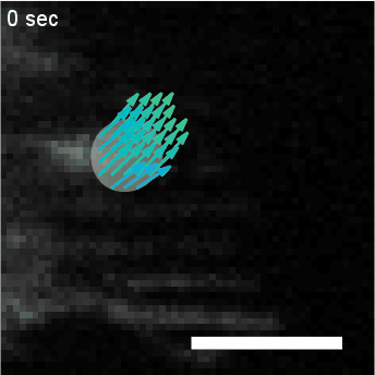
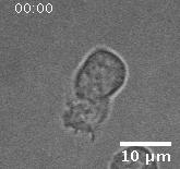
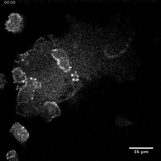
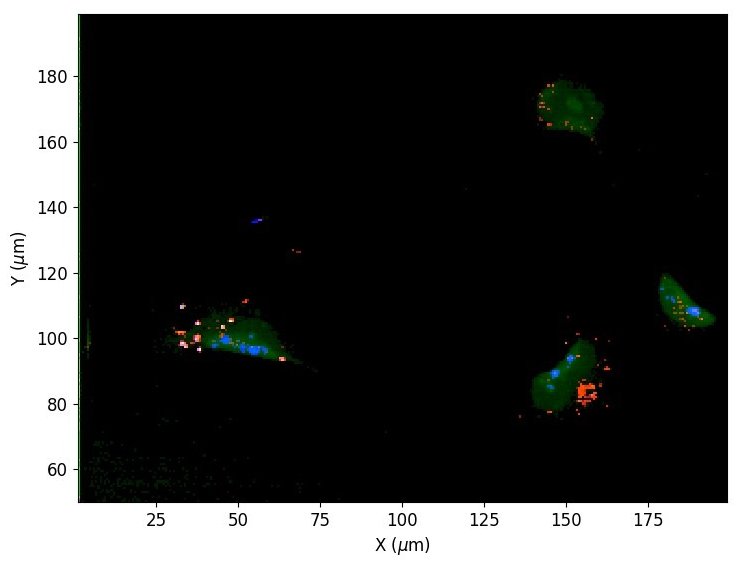
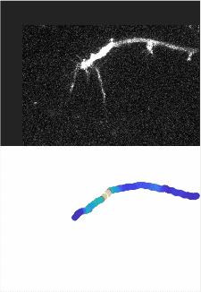
In addition to playing a role in processes such as wound healing and development, collective migration is seen in the progression of diseases such as cancer. As tumor cells become more malignant, they gain the ability to migrate throughout the body; in addition to migrating as individual cells, they have been seen to migrate collectively in vivo and there is increasing evidence that collective behavior plays a role metastsis.

Using automated image analysis techniques we are able to extract information such as velocities from images of migrating human epithelial cells. Inspired by tools developed to study fluid flows and moving grains of sand, we have quantified the motion of migrating epithelial sheets and measured differences in malignant and non-malignant cells. We are currently using these tools to understand how the motion of epithelial cells is regulated and how changes in cell migration are linked to metastatic cancer.

Publications:
R.M. Lee, D.H. Kelley, K.N. Nordstrom, N.T. Ouellette, and W. Losert, New J Phys 2013
R.M. Lee, C.H. Stuelten, C.A. Parent, W. Losert, CSPO 2016
R.M. Lee, H. Yue, W.J. Rappel, W. Losert, Interface 2017
Y. Zhang, X. Guoqing, R.M. Lee, Z. Zhu, J. Wu, S. Liao, G. Zhang, Y. Sun, et. al., Cellular and Molecular Life Sciences 2017
C.H. Stuelten, R.M. Lee, W. Losert, and C.A. Parent, Cellular Signalling 2018
R.M. Lee and W. Losert, Seminars in Cell & Developmental Biology 2019
R.M. Lee, M.I. Vitolo, W. Losert, and S.S. Martin, Scientific Reports 2021

Cell Sheet and PIV
Two images of a migrating sheet of MCF10A cells (top) compared to the velocity information derived using particle image velocimetry on these images (bottom).

Analysis of Actin Waves using Computer Vision Algorithms

Leonard Campanello, PhD Student, Physics
Matthew J. Hourwitz, PhD Student, Chemistry
with Prof. John Fourkas (University of Maryland)

Quantify the spatiotemporal dynamics of objects in biological systems often requires the analysis of diffuse concentration fields, such as fluorescent actin. In this project, we utilize a computer vision algorithm called “optical flow” to capture and coarse grain the dynamics of amorphous actin intensity fields to measure physical properties such as velocity and persistence. We use this analysis to determine the effect of surface topographies on actin dynamics, the effect of electric fields on neutrophil migration, and the ways that actin waves in Dictyostelium Discoidium cells change in response to different physical and chemical perturbations.

Publication:
R.M. Lee, L. Campanello, M.J. Hourwitz, P. Alvarez, A. Omidvar, J.T. Fourkas, W. Losert, Molecular Biology of the Cell 2020

Optical Flow of Actin
HL60 cell with fluorescently labeled actin, overlaid with clustering of optical flow vectors to quantify mesoscale actin dynamics.

Electrotaxis of Neutrophil-like Cells

Abby Bull, PhD Student, Physics
with Prof. Min Zhao (UC Davis) and Prof. Quan Qing (ASU)

Directed migration of cells is facilitated by topographic, chemotactic, and electrostatic cues during a variety of biological functions such as wound healing and immune response. It is known that in vivo electric fields attract neutrophils, and they also affect migration of many other cell types including Dictyostelium discoideum and tumorigenic cells. In this work, we combine topography and electric fields to characterize the migratory phenotypes and actin dynamics of neutrophil-like HL 60 cells in response to a variety of electrical stimuli to better understand the mechanism of electrotaxis.

Migrating HL60 cell
Migration of a neutrophil-like HL60 cell.

Spatialtemporal patterns of actin polymerization in response to electric field in oversized Dictyoslelium discodium

Qixin Yang, PhD Student, Physics
with Prof. Peter Devreotes (Johns Hopkins University)
with Prof. John Fourkas (University of Maryland)

In this study we use giant Dictyostelium discoideum (D.d.) cells to understand how actin dynamics are involved in the response of cells to external cues. Giant D.d. cells are generated by electrofusion and can be up to ten times the size of normal D.d. In the larger cells, waves of actin polymerization can travel across cell membranes and are no longer constrained by boundary effects. We use this system to study spatial-temporal pattern of actin polymerization and cellular behaviors influenced by nanotopography and electric fields.

Giant D.d. cells on a flat surface.

Developing Novel Optical Toolsets for Visualizing Cellular Dynamics Across Multiple Scales

Phillip Alvarez, PhD Student, Biophysics
Samira Aghayee, PhD Student, Biophysics
with Dr. Charles Camp (NIST), Dr. Marcus Cicerone (NIST), and Dr. Gabriel Frank (Ben-Gurion University of the Negev)

Through the use of cutting edge optical techniques such as re-scan confocal microscopy, stimulated raman scattering (SRS), and modulation of the quantum properties of light during optical stimulation, new tools being developed in Losert Lab seek to reveal the links between intracellular excitable systems such as the cytoskeleton and action potentials at both single cell and collective scales. The systems currently under development are our hybrid SRS and 2-photon imaging system and our multiscale microscope, the former of which provides for complex optical stimulation and label-free imaging of molecular targets in-vivo and in-vitro, and the latter of which provides a novel approach to simultaneously image at the collective cellular level and at the sub-cellular level beyond the diffraction limit.

BCARS image
Label-free image taken of MDA-MB-231TD cells using Broadband Coherent Anti-Stokes Raman Spectroscopy in collaboration with NIST. Image shows select Raman shifts associated with nucleotides (785 cm-1, 750 cm-1, shown in blue), proteins (1004 cm-1, 1450 cm-1, shown in green), and lipids (2850 cm-1, 1420 cm-1, shown in red).

Induced Differential Gene Expression via Nanotopographical Surfaces

Sylvester Gates, Post-Baccalaureate Faculty Assistant
with Prof. John Fourkas (University of Maryland)

Cells undergoing esotaxis on ridged nano-topographical surfaces exhibit distinct actin and migratory phenotypes compared to flat surfaces. Previous studies of gene expression have shown that electrotaxis and chemotaxis, which influence cell migration, induce changes in gene expression that differ from those in wild type or otherwise "normal" conditions. In this work, we utilize Illumina next-generation sequencing (NGS) to identify common pathways in cell migration and unique pathways that may be specific to the type of guided migration cells experience.

ProjectSchematic
By comparing different cell types both on and off ridges, gene expression changes can be correlated with exposure to nanotopographies.

Biocompatible Nano-topographical Surfaces

Matthew J. Hourwitz, PhD Student, Chemistry
with Prof. John Fourkas (University of Maryland)

Micro and nano-topographical surfaces are of great interest in our lab to probe mechanical perturbations to cell migration and actin dynamics. To fabricate these surfaces, master patterns are designed using multiphoton absorption polymerization (MAP). They can then be replicated using soft lithography and replica molding. We are working on ways to increase the scale of patterns and of output. The polymer replicas are biocompatible: we have observed overall cell survival on these surfaces for up to two months. The research also involves understanding how material and surface chemistry affect the cellular systems we probe.

Publication:
X. Sun, M.J. Hourwitz, E.M. Baker, B.U.S. Schmidt, W. Losert, J.T. Fourkas, Scientific Reports 2018

Surface Fabrication Method
Design, molding, and large-scale production of micro/nanoscale topographical patterns.

3D Reconstruction of T cell Activation Proteins

Leonard Campanello, PhD Student, Physics
with Dr. Maria Traver and Dr. Brian Schaefer (Uniformed Services University) and Dr. Hari Shroff (National Institutes of Health)

Careful regulation of T cell activation is critical to ensure that activation signals persist over controlled periods of time. If signals are too short then the immune system will not properly respond, but if the signals persist for too long then it could result in autoimmune diseases such as Type 1 Diabetes. In this project, we study the roles of several proteins in regulating T cell activation, including MALT1 and BCL10.

Structures from Image Analysis

Actin Polymerization in Axon Guidance

Leonard Campanello, PhD Student, Physics and Kate O'Neill, Post-Doctoral Research Associate
Corbett-Frank, Undergraduate Research Assistant
with Dr. Edward Giniger (National Institutes of Health)

The Abl tyrosine kinase (AKT) signaling network plays an important role in axonogenesis, regulating cell adhesion and actin polymerization. Although individual components within the signaling network are known, the relationship among them remain unresolved. We aim to reveal a subset of this multidimensional relationship: the role of regulated actin polymerization in axon guidance. To probe this relationship, we use growing axons in the wings of Drosophila as a model system. As our aim is to compute a distribution of actin concentration vs. downstream distance along these axons and study the dynamics of this distribution, we must first locate the axon within the 3D image. This project focuses on developing novel filtering algorithms and parallel-medial axis thinning to segment the axons, integrate the actin intensity along the skeleton, and produce the time-dependent actin distribution.


Axon Actin Analysis
Automatically determining the actin-intensity distribution along a growing axon in a Drosophila wing. Top: 2D max-projection of the actin-marked, Z-stacked input image. Bottom: Output of our filtering pipeline: average actin-intensity distribution along the main body of the axon.

Tracking-based Motion Correction

Samira Aghayee, Position
with Dr. Patrick Kanold (University of Maryland)

Calcium imaging provides a real time view of neuronal activity with single cell resolution. Neuronal activity is inferred from the dynamics of fluorescence signal coming from the soma membrane. However, as in vivo experiments introduce inevitable jitter to the dataset, any real-time analysis of the network's behavior requires a fast, real-time capable motion correction method. To that end we have introduced a tracking-based motion correction method that reduces the image to a set of a few central positions for the extracted bright neurons in the FOV. The neurons are tracked in time and the image sequence is then corrected to the average position. To compare the performance to the other methods in the field, a simulated dataset with known jitter was generated and the detected jitter is compared to the actual jitter.

Publication:
S. Aghayee, D.E. Winkowski, Z. Bowen, E.E. Marshall, M.J. Harrington, P.O. Kanold, and W. Losert, Frontiers in Neural Circuits 2017

Motion Correction of Neuron Position

Maryland Day: Welcome to Cells in Motion!

Lab Outreach

As part of the 2019 Maryland Day event, the Losert Lab created an interactive demonstration of our cell tracking software where participants were able to track their motion while playing games such as follow the leader. To watch our video or to learn more, visit our Maryland Day page!

MarylandDay
University of Maryland

Contact

Please contact wlosert@umd.edu for questions about the Dynamics of Complex Systems lab.